Eye examinations are kind of the bread and butter of what we do. Yes, beyond the pretty walls of our lobby, lined with hundreds of stylish frames and lens options, there are rooms and suites filled with cutting edge equipment. This equipment is all designed to aid in the examination, measuring, and diagnosing your eyes.
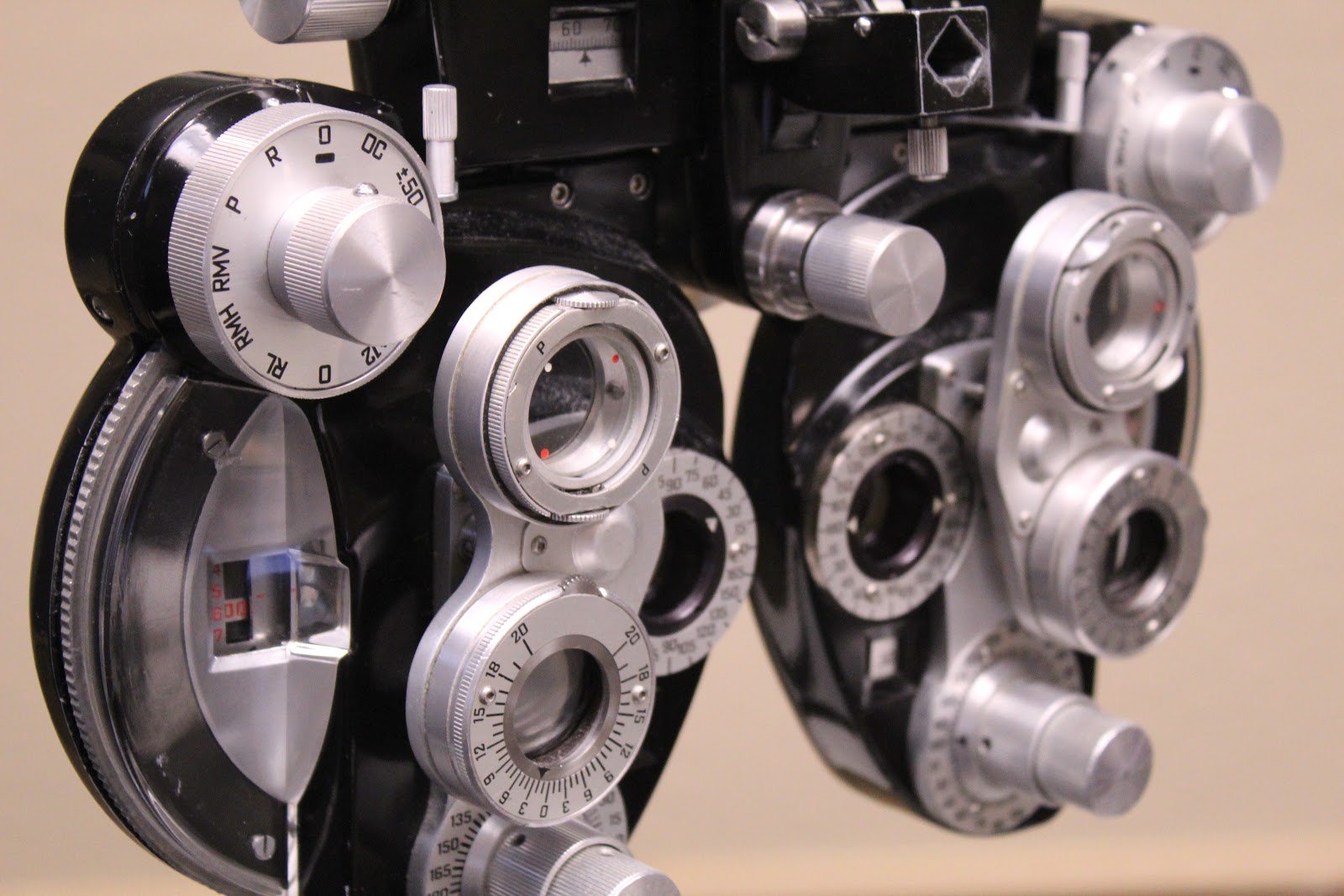
Many know the main components of an eye examination. You have the common vision chart, with its giant “E”, to roughly determine the distance you can see. Then we have color anomaly charts, introduced by Dr. Shinobu Ishihara over 100 years ago, still used today to test different color deficiencies. And we all know the goofy looking contraption with all the knobs that test lenses against each other to fine-tune your prescription (it’s called a phoropter, but we’ll go over that more later).
While most people have an idea of what all is involved in an eye examination, fewer people understand what a complete eye examination is and what’s involved.
It all begins in the lobby. While filling out the pre-appointment paperwork may seem dull and boring, it provides some important information that can be used to help determine if you are at risk or having eye issues related to a different medical condition. Prediabetic and diabetic patients, for example, would want to disclose that information so we can screen them for diabetic retinopathy or other vision complications. Another example that is equally important would be knowing what medications patients are actively taking. Patients taking high-risk medications, such as Plaquenil, need to be screened regularly for toxicity that can prevent potentially painless vision loss.
Once the paperwork is filled out and you’re called back, the real fun can begin! Believe it or not, the part of the eye exam most people are familiar with is usually towards the end of the journey. First, after you’re assessed, your retinas are examined. This can be done in one of two ways. We offer the traditional pupil dilation method that allows us to peer into your eyes, but can usually result in blurry or sensitive vision for a couple of hours after the examination. We also use a more advanced method that doesn’t require dilation and doesn’t affect your vision.
OPTOMAP
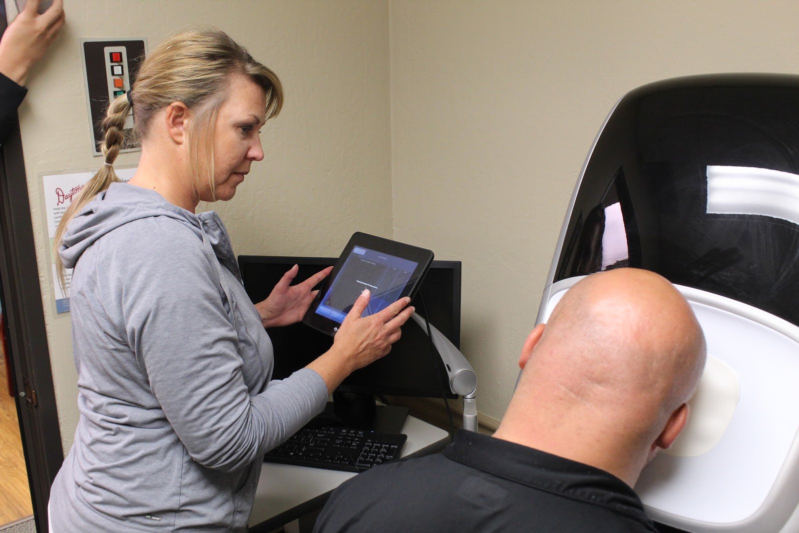
This magical, dilation-free device is known as an optomap. Our optomap consists of an array of powerful cameras that can look through our pupils and into the inner eye. With a 200 degree view, it can digitally map out your retinas and look for signs of eye diseases like retinal degenerations, retinal tears/breaks and glaucoma, to name a few. This is a major part of your eye examination. Making sure you have 20⁄20 vision is only half the battle. This step in the process allows us to look for dangers and issues noninvasively. The digital image from the optomap is saved forever so we can use it to compare this year to next year. The old saying goes, “A picture is worth a thousand words”, and that could not be more true when it comes to the eyes.

OCT
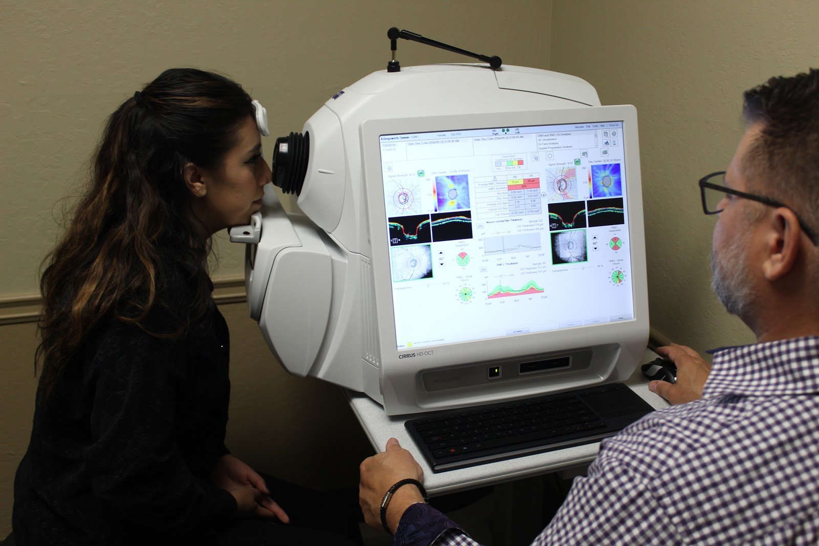
After the optomap, you’re led to a second room where you sit in front of our Optical Coherence Tomography machine (or OCT, for short). Our OCT is another set of powerful lasers that use light waves to take a cross-section picture of your retina. This cross-section can show the distinctive layers of your retina and measure their thickness, which is important in deciding and providing treatment options for retinal diseases. It can also detect certain changes in your retina that can be caused by different disorders, like glaucoma and macular degeneration.

FDT/HVFA
These first two machines are important for discovering, diagnosing, and tracking optical disorders. Once these processes are out of the way, you move on to a different set of machines that are more meant to measure certain areas of the eyes and provide critical information for prescriptions. Our Frequency Doubling Testing (FDT) Perimetry is one such machine. The FDT can be used as a basic screening to test your peripheral vision, keeping track of any vision loss around the edges of your visual field. If any significant defects appear on the screening, the Humphrey Visual Field Analyzer (HVFA) is a longer, more precise and specific test to help better quantify and localize any peripheral vision, optic nerve or neurological issues.

TOPCON AR
Next, you can move onto the autorefractor and keratometer/topographer (since ours is made by TopCon, we’ll simply call it the TopCon from now on). The TopCon machine primarily measures and maps the cornea and the estimated length of the eye to determine when your eyes can properly focus on an image. This is an important starting point for finding the right prescription for your eyewear and determining the appropriate size and shape of contact lenses. It takes a lot of the heavy lifting out of the way and can provide a relatively reliable and accurate analysis that will later be refined behind the phoropter.

VISUAL EYE TESTS AND PHOROPTER
From here, we move into the more familiar examination room. Here is where you will go through all those classic eye tests with your doctor. With them, you will be tested for things like color anomalies/deficiencies, depth perception, and visual accuracy. After these tests, you’ll look through the many lenses of the phoropter. Phoropters are used to fine tune your prescription, making sure your lenses won’t cause strain or headaches after long uses. An accurate prescription is critical, so your doctor will guide you through a series of simple questions like “which is better, 1 or 2?”

REVIEWING THE IMAGES/FINDINGS
Your doctor will then review with you images taken during pre-testing. He/she will discuss the results of your exam pertaining to your visual correction and health of the eyes including critical findings, treatment/management, and follow-up recommendations.
TRYING ON FRAMES
Finally, the examination comes to an end. A full assessment has been made and you can begin trying on frames that match your style. With hundreds of options to choose from and our brilliant opticians there to help you, you’ll be guided through the different options we offer for frames and lenses alike. Lastly, we prepare you to come back next year so we can do it all over again even if you don’t need glasses, we need to make sure the health of your eyes are good.


